LISFRANC (Tarsal-Metatarsal) FRACTURE DISLOCATION
The Lisfranc injury to the forefoot is thought to have originated with Napoleon's surgeon. During that time, if a rider was thrown from his horse and the foot was caught in the stirrups, the rider often suffered a severe fracture of multiple bones in the midfoot region with dislocation of the fragments. The resulting trauma often resulted in an amputation of the forefoot at a specific level called the Lisfranc joint, after the French surgeon who first noticed the pattern of this injury. Fortunately, although the injury is still called a Lisfranc injury, amputations are rarely done for this injury.
ANATOMY
However, the term Lisfranc injury (or fracture-dislocation) now defines a specific type of injury to the forefoot. To understand the injury it is first necessary to understand the anatomy of the foot. The talus is the bone below the ankle, which allows for the upward and downward motion of the ankle. Beneath the talus is the calcaneus, which is the heel bone. Just in front of the talus (when viewed from the top (dorsal) aspect of the foot, is the navicular bone. Just beyond this bone, towards the toes are the bones that are responsible for the arch configuration of the midfoot.
The bones consist of the 3 cuneiform bones and the cuboid bone. Further towards the toes, are the metatarsal bones. Everywhere that these bones come together, there is a joint. The proper alignment of the bones at the joints is due to the ligaments that hold the bones in the proper place. The Lisfranc joint actually refers to a number of joints which are formed by the junction of the metatarsals and the cuneiforms, and by the junction of the metatarsals and the cuboid bone.
INJURY & DIAGNOSIS
In modern times, a Lisfranc injury indicates an injury to the normal alignment of the cuneiforms and metatarsal joints with the loss of their normal spatial relationships.
The most common injury to the Lisfranc joint occurs at the joint involving the 1st and 2nd metatarsals and the medial cuneiform. If the ligaments between the medial and mid cuneiforms are disrupted, or between the 1st, 2nd mt and the medial cuneiform, then the bones separate and the normal alignment of the joints is lost.
When recognized, this injury may be treated surgically and has a much better prognosis then when it is not diagnosed.
Obviously, with multiple fractures, injury to the Lisfranc joint is obvious. However, the difficulty develops with the subtle injuries that may result in a small chip fracture, or simply a torn ligament. Unfortunately, these injuries are difficult to diagnosis. If not treated, chronic pain often results.
Accurate diagnosis requires a physical exam to determine the amount of swelling, the location of the bruising, and the areas of tenderness. When the injury is suspected, x-rays are often taken of the foot. However, due to the subtle nature of the changes associated with this injury, multiple films are often necessary. In addition, even if the first set of x-rays is normal, as the swelling decrease with time, this may allow for the bones to move from their normal position, especially if the stabilizing ligaments have been torn and the joints have been made unstable. For that reason, it is often necessary to take x-rays during the healing process to evaluate for the possible delayed development of instability. In some patients, it is necessary to proceed to more sophisticated scanning techniques including CT scans or MRI. Unfortunately, it is possible to have a scan that is interpreted as normal, even when the joints are unstable and partially dislocated. (This is referred to as a false negative finding).
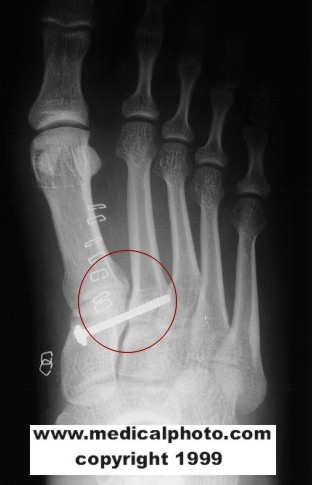
TREATMENT
Once instability at the midfoot joints has been established. The treatment is usually surgical if a significant separation of the bones exists. Surgical treatment usually requires that pins and/or screws be inserted to stabilize the bones and joints. This procedure is called an open reduction and internal fixation. This allows the normal anatomy to be re-established.
After surgery, it is critical that the patient remain non-weight bearing until full healing occurs. During this time, a short leg cast is usually applied. Once healing has occurred, several months after the surgery, it is necessary to remove the screws/ pins (internal fixation devices) prior to full weight bearing and resumption of activities.
THIS MATERIAL DOES NOT CONSTITUTE MEDICAL ADVICE. IT IS INTENDED FOR INFORMATIONAL PURPOSES ONLY. PLEASE CONSULT A PHYSICIAN FOR SPECIFIC TREATMENT RECOMMENDATIONS.
THE CENTER FOR ORTHOPAEDICS AND SPORTS MEDICINE
1211 JOHNSON FERRY RD.; MARIETTA, GA., 30068
770-565-0011
Copyright ©2000 by The Center for Orthopaedics & Sports Medicine - ALL RIGHTS RESERVED
No comments:
Post a Comment